Chemical biology is an interdisciplinary field that combines principles and techniques from both chemistry and biology to study and manipulate biological systems. It aims to use chemical tools and approaches to understand the complex processes that occur within living organisms at the molecular level.
The following list profiles the current research being conducted at the Kikuchi Lab
| Design and synthesis of sensor molecules for visualizing biological events in living cells | Design and synthesis of sensor molecules for visualizing dynamic movement in animals | Design and synthesis of sensor molecules for regulating the function of proteins in living cells | Development of chemical techniques for modification and labeling of proteins in living cells | Development of a novel method for posttranslational knockdown of a protein function |
The crucial aspect in chemical biology research is whether the developed chemical tools can truly be applied to biological studies. In our laboratory, we aim to create new research materials by setting the hurdle of whether they can genuinely be applied to biology as a criterion for our research content.
One of the prominent trends in recent scientific research is interdisciplinary studies at fields such as physics, chemistry, and biology, breaking down traditional boundaries. In recent years, advances in life science have been leading the overall scientific research. Our laboratory aims to engage in life science research with chemistry as its foundation. We provide research environment that is optimally designed to promote interdisciplinary research spanning the fields of physics, chemistry, and biology. While the fundamental discipline in our laboratory is chemistry, we also conduct studies in biology and physics as needed. In other words, by keeping an eye on applicability, we believe that students will acquire a broad range of knowledge and experience that extends across multiple fields. In our laboratory, we consider research as a platform for self-expression. In other words, we believe the essence of research life lies in demonstrating your original ideas by conducting various kinds of experiments. There is no greater joy than finding out that your research has real outcomes. Of course, success is not always guaranteed, but it makes research more fantastic. It is important to think positively, not to be disappointed, and carry on. With careful consideration and effort, great results will undoubtedly come. If you have an interest in life science research using chemistry, we warmly invite you to knock on our door.

Due to the advancement of biochemistry and genome decoding, a multitude of signaling molecules and their receptors within living organisms have been identified one after another. The term "post-genome" is now commonly used in this era, where the elucidation of functionality under physiological conditions is emphasized as the next goal. In previous biology research, the functions of biomolecules were examined by sacrificing animals or cell disruption. On the other hand, it is believed that more information can be obtained if it were possible to study the functions of biomolecules while the animals or cells are still alive. Therefore, we are tackling this problem using the approach of Chemical Biology. Until now, there were very few probe molecules available to capture the reactions of biomolecules at a practical level, and this has become a subject of further investigation. We are designing and synthesizing probe molecules that react specifically with and visualize intracellular molecules, and directly applying them to biological research. In other words, we design molecules that change the intensity of fluorescence or color when they encounter intracellular molecules, or molecules that alter MRI contrast.
Specific Research Topics
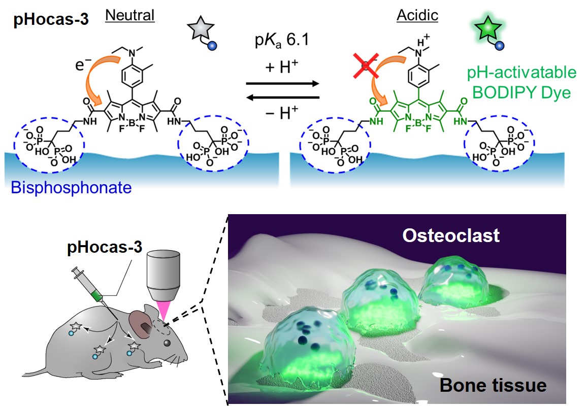
Visualisation of cellular functions in vivo is an crucial technology that not only pertains to fundamental research but also holds significant implications for medical applications like gene therapy. However, there are very few practical probes available for detecting molecules associated with these cellular functions within live organisms, and their development is highly anticipated. We have, thus far, developed a fluorescent probe capable of detecting the activity of bone-dissolving cells (osteoclasts) within living organisms and clarified their functionality. The abnormal activity of osteoclasts in bone tissue is known to lead to diseases such as osteoporosis. Until now, osteoclast activity has been examined by culturing osteoclasts on dentin slices and measuring their dissolved dents. However, this method doesn't allow for time-lapse and quantitative analysis. Therefore, we have developed a fluorescent probe that enables direct imaging of osteoclast activity in vivo (Fig. 1). Since activated osteoclasts dissolve bone by releasing acid, we chose a fluorescent dye that shows fluorescence when it comes to the acidic environment. In addition, a bisphosphonate group has been introduced to the fluorescent dye in order to deliver it to bone tissue. Thus, it selectively tells you the areas of bone that are being dissolved by osteoclasts. We injected the fluorescent probe into mice and observed using a two-photon excitation microscope, which can observe the inside of the bone. Then fluorescent signals were observed between the osteoclasts and the bone surface, allowing detection of the area of the bone that was being dissolved. By using a dye with high photo-stability, we have succeeded to observe real-time bone dissolution in accordance with osteoclast mobility. Furthermore, quantitative evaluation of osteoclast activity is possible. It is expected to lead to the development of therapeutic drugs for osteoclast-related diseases such as osteoporosis and rheumatoid arthritis.


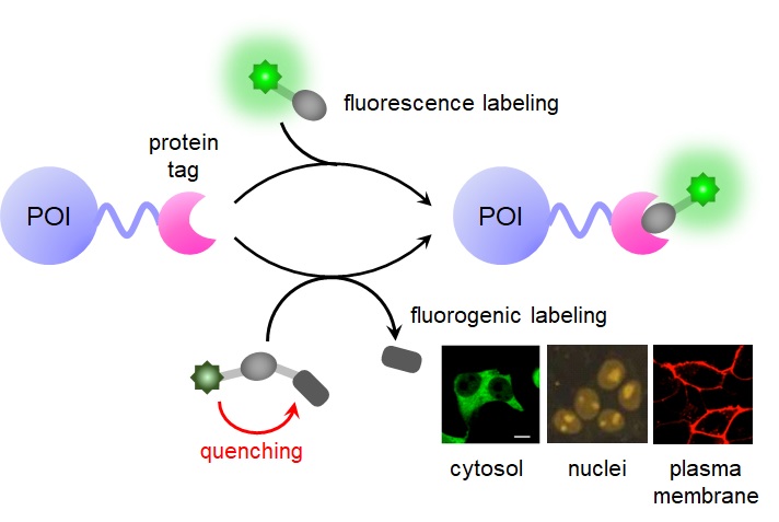
Real-time visualisation of protein localisation, dynamics, and interactions in vivo is considered an important way to elucidate their functions. A representative example of this technology is using fluorescent proteins which is the subject of the Nobel Prize in 2008. Fluorescent proteins emit fluorescence on their own. By connecting fluorescent proteins to the protein of interest (POI), the functions of various proteins in vivo have been visualized and clarified. However, fluorescent proteins have some weaknesses; being unable to control the timing and amount of labelling. Therefore, as an alternative to fluorescent proteins, we are developing a fluorescent labelling technique using fluorescent probes and proteins that specifically bind to each other (Fig. 2). In this fluorescent labelling method, a tag protein is fused to the POI and expressed in the cell by use of genetic engineering. Then the tag protein is labelled with a fluorescent probe that binds specifically to the tag protein, and in the end, we detect the fused target protein. Recently, tag protein labelling has been attracted because it has been shown to be useful in cutting-edge imaging methods such as single-molecule imaging and super-resolution imaging.

We have further developed this method and found a principle in which fluorescence is emitted only when the tag protein binds to the probe (fluorogenic-type labelling method). In this method, we are easily able to label and identify the POI without washing off the probe. We have developed two fluorogenic-type libelling methods (Fig. 3). One is BL-tag which is the mutant of β-lactamase from bacteria and the other is photoactive yellow protein (PYP) from red sulphur bacteria. In the way using the BL-tag, a quencher that quenches the fluorescence of the fluorophore is introduced into the probe. This quenching is caused by the mechanism of fluorescence resonance energy transfer (FRET). When the compound is labelled with the BL-tag, the quencher dissociates and the fluorescence intensity increases. In the other method using the PYP tag, it is based on the principle of association quenching and dissociation of fluorescent dye, so the fluorescence intensity increases when the probe binds to the tag. In fact, it has been shown that the fluorescence intensity increases rapidly, binding the tag protein with the fluorescent probe. In addition, we have already used it successfully for fluorescence imaging of proteins expressed locally in the cell membrane and inside of the cell. Fluorogenic-type libelling method is a powerful technique for the Spatio-temporal analysis of protein dynamics in detail. First in the world, we have applied this technique to reveal the role of glycan in the membrane translocation of GLUT4, a protein involved in type II diabetes and the dynamics of DNA methylation involved in gene expression. For the development of life science study, we are now applying these labelling methods to a variety of proteins and fluorescent dyes.

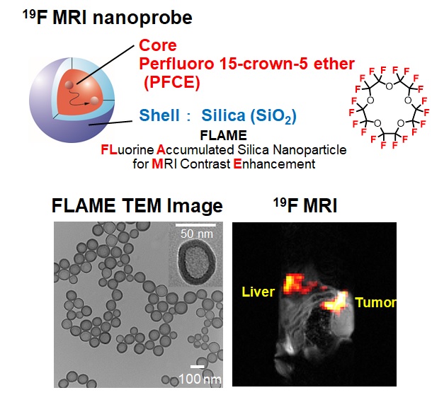
Magnetic resonance imaging (MRI) is a medical imaging technique used to visualize internal structures of the body in detail. It is one of the most clinically applied medical imaging technique benefiting from is excellent tissue resolution, nonradioactivness and non-invasiveness. In clinical settings, the commonly used MRI technique utilizes the observation nucleus as 1H (hydrogen), referred to as 1H MRI. However, our research focuses on the observation of a heteronuclear 19F. Since only trace amount of 19F exist in the body, using it as the observation nucleus in MRI allows for obtaining high-contrast images without background signals. Therefore, it is believed to be suitable for tracking enzyme reactions deep within living organisms. As an example, we have successfully created a probe for detecting the activity of caspases, enzymes involved in the regulation of apoptosis, a form of cell death. To date, we have developed the mechanism of changing 19F signal in response to enzymatic reactions by utilizing the paramagnetic interaction of Gd3+ complexes, and have designed and synthesized probes based on the mechanism. As an example, we have succeeded in developing a probe that detects the activity of caspase; an enzyme involved in the regulation of apoptosis. We synthesized a probe by attaching a Gd3+ complex that quenches the 19F MRI signal via paramagnetic interaction through a peptide containing a caspase cleavage sequence to a 19F compound (Figure 4). The probe is designed so that when the peptide is cleaved by the enzymatic reaction of the caspase, the Gd3+ complex dissociates from the 19F compound and the 19F MRI signal is turned on. Upon actual MRI imaging, we confirmed an increase in contrast associated with the enzyme reaction.

Therefore, we developed nanoparticles containing fluorine compounds, named FLAME (FLuorine Accumulated silica nanoparticle for MRI contrast Enhancement) (Figure 5). These nanoparticles have a structure where a liquid containing perfluorocarbons (PFC), a type of fluorine compound, is encapsulated within a silica (SiO2) shell. Since the PFC does not leak out from the silica shell, it allows us to detect signals derived from numerous fluorine atoms contained inside. We created a nanoparticle-type 19F MRI probe by attaching a Gd3+ complex to the surface of FLAME through a peptide containing a caspase cleavage sequence. Similar to small molecules, the 19F MRI probe we developed shows signal recovery upon cleavage. With this 19F MRI probe, we achieved the detection of caspase activity within the body of a mouse. Moreover, by altering the encapsulated fluorine compounds, it is also possible to track the in vivo dynamics of the nanoparticle-type 19F MRI probe in multiple colors. These achievements can provide the foundational technology for visualizing biological signals using MRI. As a result, we were able to visualize the localization of FLAME within the living body of a mouse and successfully image the signals of FLAME incorporated into cancer tissue. Furthermore, the surface of silica can be easily modified by functional molecules. We have prepared a nanoparticle-type 19F MRI probe in which a Gd3+ complex is connected to the surface of FLAME via a peptide with a caspase-cleavage sequence. Similar to small molecules, the 19F MRI nanoprobe we developed shows signal recovery upon cleavage. It is also possible to do the 19F MRI multicolour imaging in the body by changing the PFCs content. These results provide us with a fundamental technology for visualizing biological signals by MRI.

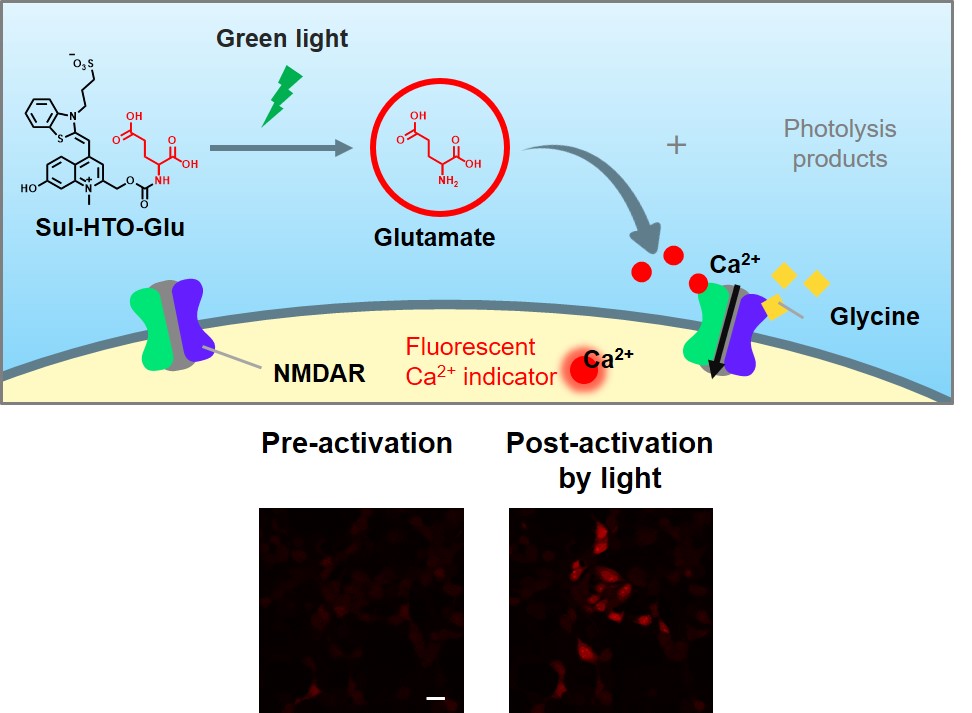
We are also advancing research that utilize chemical reactions triggered by light irradiation and apply these reactions to fluorescent imaging and the release of bioactive substances. Caged compounds are molecules where bioactive substances or drugs are protected by photolabile protecting groups. These molecules can be released at specific location and timing upon light irradiation, enabling the control of cellular functions. We have reported the release of drugs using caged membrane-disrupting peptides and the control of transcription factor function using caged ligands. While most caged compounds require ultraviolet light for molecular release, we have recently developed new protecting groups that are cleaved by visible light. We are also developing fluorescent molecules with photo-switching ability, which spectral properties are reversibly changed by light irradiation.